Guided Surgery
Guided Surgery
Guided dental implant surgery utilizes advanced Cone Beam 3D imaging to place implants at exact specifications. These plans can be implemented in real-time and setup accordingly to exact measurements.
Benefits for dental implant patients
- Precise implant placement
- Less risk for complications
- Faster and less invasive implant surgery
- High quality restorations with exact specifications
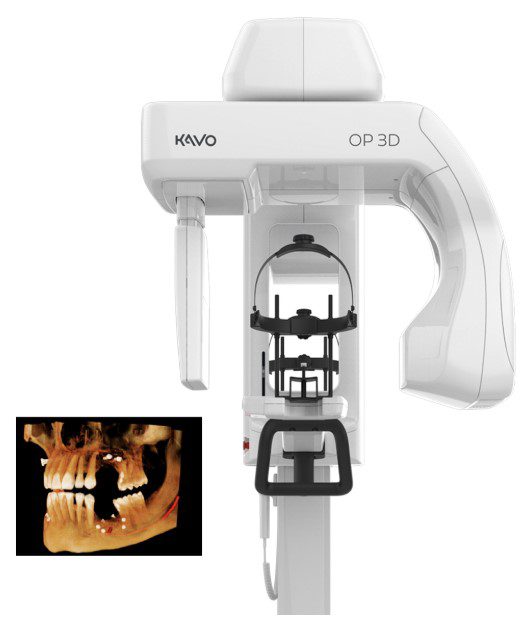
Dental cone beam computed tomography (CT) is a special type of x-ray used in situations where traditional dental x-rays just aren’t enough. It isn’t used regularly because the radiation exposure from CT scanners is greater than that of regular dental x-rays. The CT scanner uses advanced technology that generate three dimensional (3-D) images of the mouth in a single scan. The scan captures dental structures, soft tissues, nerve paths, and bone in the face. Images captured with our cone beam CT allow for more accurate treatment planning.
CT scanners are effective for:
- Precise surgical placement for dental implants
- Surgical planning of impacted teeth (wisdom teeth)
- Diagnosing temporomandibular joint disorder (TMJ)
- Evaluation of the jaw, sinuses, nerve canals, and nasal cavity
- Locating the origin of pain or pathology
- Cephalometric analysis
- Reconstructive surgery
- The focused x-ray beam reduces scatter radiation, resulting in better image quality.
- A single scan produces a wide variety of views and angles that can be manipulated to provide a more complete evaluation.
- Cone beam CT scans provide more information than conventional dental x-ray, allowing for more precise treatment planning.
- CT scanning is painless, noninvasive and accurate.
- A major advantage of CT is its ability to image bone and soft tissue at the same time..
- No radiation remains in a patient’s body after a CT examination..
- X-rays used in CT scans should have no immediate side effects